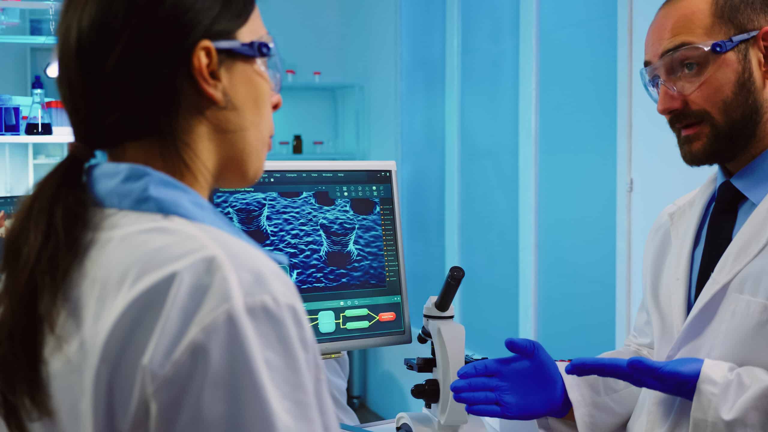
Understanding cellular functions like migration, morphology, and physiological changes in healthy and diseased cells is important for grasping biological processes and using them to optimize the creation of biological therapeutics. Live-cell imaging provides an instant picture of a cell, tissue, or organism’s current state, as close to in vivo as possible, without introducing artifacts seen with fixed cells.
Cell imaging is a noninvasive way of tracking health-related attributes and disease-related abnormalities in cancer and other disorders. Cell imaging presents important information about the etiology, progression, and ultimate fate of diseases. Imaging can automatically quantify the properties of cells, and profile phenotypic changes that can occur during disease, growth, and genetic modification of cells. Live imaging is crucial for understanding cellular functions and processes because it allows the accurate, and often real-time, reconstruction of cellular events. It is an advanced over-fixed cell observation that only provides a single point in time and not the entire process a cell goes through. However, it is also necessary that live cell imaging techniques provide 100% penetration. Missing a step or process can mean overlooking a key event in healthy or diseased cells.
Types of Cell Imaging Techniques
Light microscopy
Brightfield Microscopy — The most traditional (and oldest) form of microscopy, this technique remains valuable for conducting label-free cellular assays to look at cell number, proliferation, health, confluence, and cytotoxicity. Based on changes in light absorption, refractive index, or color to generate contrast, this technique remains a favorite optical microscopy technique. As light passes through a cell (or tissue or organism), regions altering light wave direction, speed, or spectrum generate contrasts when light rays are gathered and focused.
Phase Contrast Microscopy — This technique translates tiny variations in phase into corresponding changes in light amplitude, which are then visualized as differences in contrast in the image. A specialized condenser with annuli matches a set of objectives containing rings in the back focal plane. This technique can increase contrast when imaging living cells in culture. However, it can also produce excessive halos on the outlines of edges, reducing the visibility of boundary details.
Differential Interference Contrast (DIC) Microscopy — With this technique, plane-polarized light, and light-shearing prisms exaggerate tiny differences in specimen thicknesses and their refractive indexes. This enables better images of cellular lipid bilayers because of refractive differences between aqueous and lipid parts of the cell.
Fluorescence Microscopy
Basic Fluorescence Microscopy — This still involves an optical microscope, but one that uses fluorescence instead of scattered or reflected light to observe cell structure and functions. With this technique, a shorter wavelength of light excites a fluorophore, which then emits light at a longer wavelength. This light is detected by the microscope.
FMM/BMI/SIMI — The backbone of the Halo Labs Aura system of analyzers, this technology combines the effects of existing light and fluorescence microscopy. Backgrounded membrane imaging (BMI) is a high-contrast imaging technique involving the use of a high-contrast membrane that develops clear pictures of particles in a sample. It only requires 5 µl of sample and can deliver results in one minute. Fluorescence membrane microscopy (FMM) labels particles using specific fluorescent dyes or antibodies, resulting in simple and definitive identification. SIMI, or side illumination membrane imaging, works with BMI and FMM by detecting light scattering, specifically high refractive index particles, including those protruding out of the membrane surface. Particles that scatter light with SIMI indicate inorganic matter protruding out of the membrane such as glass or plastic. Particles that absorb light are indicative of metallic particles, such as residual Dynabeads™️, and absorbing oils that also protrude from the membrane.
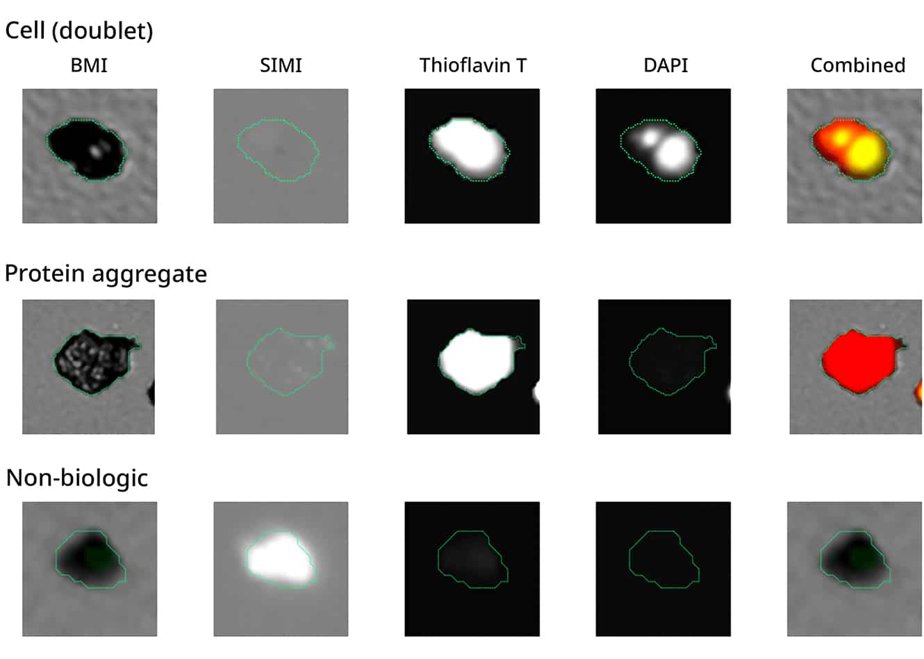
Advanced particle analysis is possible with the combination of BMI, SIMI, and FMM (Thioflavin T & DAPI) tools.
Confocal Microscopy — A type of fluorescence microscopy that uses certain optical components to generate high-resolution images of material stained with fluorescent probes. This method is capable of generating sharp, three-dimensional images. It uses a spatial pinhole to block out-of-focus light in image formation.
Two-Photon Microscopy — Also called 2PEF or TPEF, this type of microscopy is a little different from standard fluorescence microscopy because it is based on simultaneous excitation by two photons with longer wavelengths than the emitted light. The laser focuses on a specific location in the tissue and scans across the sample to sequentially produce the image. This technique is useful for tissue up to 1 mm thick.
Advanced Techniques
Super-resolution (STED, PALM, STORM) — This technique generates images of much higher resolution than light microscopy, which is limited to about 200 nm. Stimulated emission depletion (STED), stochastic optical reconstruction microscopy (STORM), and photoactivation localization microscopy (PALM) can provide resolutions about 20 times greater than light microscopes.
Total Internal Reflection Fluorescence (TIRM) — This method tracks and detects objects by using light scattered from an oscillating wave field near a dielectric interface. It has a high signal-to-noise ratio and a high vertical spatial resolution.
Electron Microscopy — These microscopes focus beams of electrons instead of photons to produce an image of a specimen. They are regarded as higher resolution and as having greater magnification than light-based microscopes.
Transmission Electron Microscopy (TEM) — Used to image the smallest structures in matter, TEM uses a beam of electrons that are transmitted through a specimen, revealing the internal organization and composition of a tissue, cell, or other material.
Scanning Electron Microscopy (SEM) — Like other electron microscopy methods, SEM uses beams of electrons, but SEM looks at the surface and composition of a sample, rather than internally.
Other Imaging Techniques
Atomic Force Microscopy (AFM) — This technique can develop nanoscale images of the surfaces of biological materials in their native environment. With this technique, a mechanical probe uses electric forces to generate tiny but accurate and precise movements on (electronic) commands for precise scanning.
High-Content Screening — This encompasses several assays, often using fluorescent dyes, to visualize cells and determine functions and operations within cells. The resolution of high-content microscopy techniques has surpassed that of traditional microscopy, and these techniques incorporate automated imaging and analysis for large-scale studies of cells.
Cell Imaging Data Analysis
Image Processing — There are several techniques available for enhancing image quality and clarity. In addition, scientific organizations have developed checklists for publishing high-quality, clear microscopic images. First, in what may seem an obvious step, be sure that your microscope is properly aligned—condenser and lens—and use the correct diaphragm setting (for light microscopy). Guidelines are also available for other methods, such as fluorescence microscopy.
Christian Tischer, a team leader in the European Molecular Biology Laboratory (EMBL) Data Science Centre, emphasized that “for microscopy-based research, this ranges from issues like the legibility of image data in publication figures, providing scale information, and a responsible choice of contrast adjustments, to sharing image data on public archives and making accessible the analysis pipeline on cloud computing platforms.”
Halo Labs Aura system of particle analyzers combine light (i.e., brightfield) and fluorescence technologies to create higher sensitive assays with higher resolution images. The system also includes Particle Vue analytical programs that help improve images and provide quantitative, workable data.
Quantitative Analysis — Images don’t serve much of a purpose if quantitative data can’t be extracted from them. Several tools can count cells and particles in a sample, assess cytotoxicity with live/dead assays, measure autophagy, determine cell phenotypes such as cell shape, size, and structure, locate specific molecules in cells, and analyze dynamic processes over time.
Data Interpretation
Morphological Analysis — Determining the size, shape, and structure of cells and other particles and components in culture can help evaluate the effectiveness of treatment candidates, as well as illustrate changes that occur due to disease. The Aura family of analyzers and Particle Vue analytical software can accurately determine morphology and changes that occur in healthy and diseased cells and in reactions to drug treatments. A common method to determine viable vs. non-viable cells is staining the cells with DAPI and using FMM on Aura to determine the population of cell conditions.
Molecular Localization — Pinpointing the exact location of specific molecules within cells can help determine their role in health, disease, and reactions to treatments. These techniques, such as Single Particle Tracking (SPT) and Single Molecule Localization Microscopy (SMLM), can identify molecules on a nanoscale, and determine the positions and movement of proteins and other important molecules within a cell.
Time-Lapse Imaging — Monitoring cellular responses over a period of time can help determine the right time to read endpoint assays, characterize cellular kinetics of multiple reactions at once, and track real-time effects of treatments on cells. Automated time-lapse imaging can dramatically reduce analysis times, as well.
Advanced Analytical Techniques
3D Reconstruction — These techniques take imaging data and create three-dimensional models of cells and cellular components. 3D cell culture (or automated construction) provides more relevant physiology than traditional monolayer (2D) techniques.
Fluorescence Lifetime Imaging (FLIM) — This advanced technique allows for simultaneous recording of the fluorescence lifetime and spatial location of fluorophores in an image. FLIM can help researchers investigate pH, ion concentrations, solvent polarity, non-covalent interactions, viscosity, and oxygen tension in live cells.
Applications of Cell Imaging Techniques
Cell Biology Research
Studying Cell Structure and Function — The structure of cellular organelles and membranes is key to understanding how cells develop and how genetic, physiological, and development processes occur. This can be important for basic comprehension of how cells work, but also how they behave in diseased states. Imaging can also help determine the function of these cellular structures. Cell imaging in time-lapse can illustrate what changes these structures go through (or activate) in development, health, disease, and death.
Tracking Cellular Dynamics — Imaging techniques help researchers observe cell migration, division, and differentiation, all in real-time. Cellular processes span multiple time and length scales, including developmental processes and cancer metastasis. Different techniques can determine how these processes work, either in real-time, in three dimensions, or in cellular/subcellular membranes.
Medical Research
Disease Mechanisms — Cell imaging techniques provide a clearer picture of how diseases arise in a cell, take over cell function (in some instances), and ultimately can damage or kill a cell. Three-D microscopy is being studied for the possibility of seeing nerve cell changes during Alzheimer’s or Parkinson’s disease, while super-resolution microscopy is used to visualize and quantify platelet membrane proteins during immune responses. Other fluorescence techniques are being used to visualize how CAR T-cell therapies work and to better understand controlled cell death.
Drug Discovery and Development — Getting accurate and detailed images of cell structure and function in reaction to drug candidate molecules is a crucial part of discovering and developing new therapies and vaccines for disease. Techniques like the Aura system of particle analyzers can use FMM and BMI, combining light and fluorescent microscopic methods to determine how cells respond to drug candidates and what other molecular and structural factors may be interfering with that response.
Genetic Research
Gene Expression Studies — Matched with sophisticated genetics techniques such as next-generation sequencing, microscopy can identify the phenotype changes that occur with changes in genotype. Electron microscopy can identify specific proteins and other molecular changes, while light and fluorescence microscopic techniques can produce images of how cellular structures and functions change in reaction to genetic alterations. Advances in live-cell imaging allow for the visualization of gene expression and intracellular interactions, in real-time.
CRISPR and Genetic Engineering — CRISPR-Cas9 is a powerful and versatile gene-editing tool, but it also can be used to visualize DNA replication and mutations in live cells. The Cas9 protein, labeled with a fluorescent protein, can label a targeted locus and be visualized using microscopy. Other scientists are looking at how CRISPR can develop three-dimensional images of the genome and portraits of entire chromosomes. Likewise, molecular imaging in reporting and assessing the status of cell grafts – as well as their relation to the local microenvironment, cell replacements, and/or gene replacements – makes cell imaging a promising way to determine the effects of genetic engineering techniques.
Conclusion
While imaging has made dramatic advances even in the last few decades, observing cells and subcellular processes at high resolution while maintaining environmental context still presents challenges, especially in a minimally invasive fashion. Cellular resolution, especially in context and in living organisms, still needs more development (several experimental projects are underway). Observing proteins in cells – including their structure, quantity, location within cells, and activity – can be done with electron microscopy but techniques that can see in real-time and that can see smaller molecules are needed. Data collection and analytics need more universal standards, and better global access to these technologies will dramatically advance scientific research.
External references
https://www.nature.com/articles/s41592-023-01987-9
https://www.nature.com/articles/s41592-021-01156-w?fromPaywallRec=false
https://www.nature.com/articles/d42473-022-00356-y
DISCOVER THE AURA FAMILY


Read More ON this topic
")


